
Tuesday, 29 March 2011
An easy/cheap/high-throughput screening method for NHase... not yet
I see there is a new paper coming out in the Journal of Microbiological Methods by Zheng and co-workers at Zhejiang University of Technology with the rather interesting title of "Ferrous and Ferric ions-based high-throughput screening strategy for nitrile hydratase and nitrilase" [doi:10.1016/j.mimet.2011.03.005]. Sadly the paper doesnt quite live up to that general title since it relates a nice way of following these activities but only on alpha amino nitriles as starting material. Finding a general way to track nitrile conversion to amide is something that doesnt have a current low tech answer (and Zheng give a good summary of the current best of breed examples out there, both those with rather specific substrate requirements and those requiring a more instrumenttal approach). In the end, I guess, you can always rack and stack your samples on a HPLC or LCMS, and let the robot churn through...

Friday, 11 March 2011
NHases in organisms that aren't bacterial
A protein search amongst NCBI or SwissProt's data for NHases that arent in bacteria has been a pretty fruitless task. There are two notable exceptions to the "NHases are in bacteria, period" rule. The paper [Foerstner KU, Doerks T, Muller J, Raes J, Bork P. A nitrile hydratase in the eukaryote Monosiga brevicollis. PLoS ONE 2008;3:e3976.] showed that there is something in an aquatic metazoan Monsiga brevicollis which is combines the alpha and beta subunit in one protein chain. That sounded interesting so we have done some molecular biology magic and put that sequence in E. coli, and it expresses OK, though we havent had the time/manpower to play with it yet. The other outlier is pointed out in Prasad and Balla's Biotech Advances review of last year which points out that the plant Ricinus communis has also got the primary sequence for a NHasetoo. The usual tag sequence (VCTLCSC) in the alpha subunit suggests it is a cobalt centred one and if you ClustalW that with the cobalt-centred NHase which is featured in pdb file 1V29 (from Bacillus smithii) you get an alignment score of 54. Interesting.


Tuesday, 8 March 2011
ClustalW analysis of pdb sequences
Following on from looking at the similarity of the pdb files, I thought I would run a ClustalW analysis of the primary sequences I used for that analysis. A lot are very similar (>98%) as you might expect, and once again it is obvious these files actually represent one iron centred and two cobalt centred templates. The iron centred template has scores between 40 and 50% for the cobalt centred pair, and the score between the two cobalt centred templates is about 60%.
Monday, 7 March 2011
How many clearly different structures of NHase are reported as pdb files?
Another helpful thing that the Iterative Magic Fit function within DeepView can do is provide a numerical value (based on a RMS calculation) for the similarity of the three dimensionality of the enzymes being overlaid. After concentrating on enzymes which are clearly NHases, discarding structures which are clearly mutants of an existing structure (which is either obvious from the title or the combination of author and date of submission), you get left with 13 distinct pdb files [1AHJ, 1IRE, 1UGP, 1V29, 2AHJ, 2CYZ, 2CZ6, 2D0Q, 2DPP, 2QDY, 2ZPB, 3A8O and 3HHT]. I then IMFed every one of these against each other to get a matrix of values for similarity. Basically if they had a value of less than 0.5, they look pretty much the same, greater than 1 than they look noticeably different.
The results of this showed that ALL the iron centred enzymes have RMS of <0.5 with each other which is not entirely surprising since they are all from Rhodococcus species. They all have RMS of greater than 1 for all the cobalt centred enzymes. There are two distinctly different cobalt centred arrangements: the Pseudonocardia thermophila pair of 1IRE and IUGP, and the trio of Bacillus structures of 1V29, 2DPP and 3HHT. Within these groupings they have RMS of less than 0.5, and between the groupings RMS of greater than 1.
Effectively this says to me that there are three basic structures for NHase known currently- one iron based and two cobalt based. Below is an excerpt from a table holding all the results from these calculations which shows this clustering.
PDB ID | ||
1.06 | ||
1.06 | ||
1.06 | 0.07 | |
1.19 | 1.14 | |
0.45 | 1.09 | |
0.44 | 1.07 | |
0.47 | 1.09 | |
0.45 | 1.09 | |
1.22 | 1.16 | |
0.46 | 1.09 | |
0.44 | 1.08 | |
3A8O | 0.47 | 1.11 |
3HHT | 1.22 | 1.15 |
Wednesday, 2 March 2011
NCBI- March numbers for NHase
A quick text search for "nitrile hydratase" under proteins on NCBI's website shows 2677 (+5 since the start of February) hits for the phrase, and 988 (+3 since the start of February) RefSeq hits. The number of PDB hits is unchanged at 36.
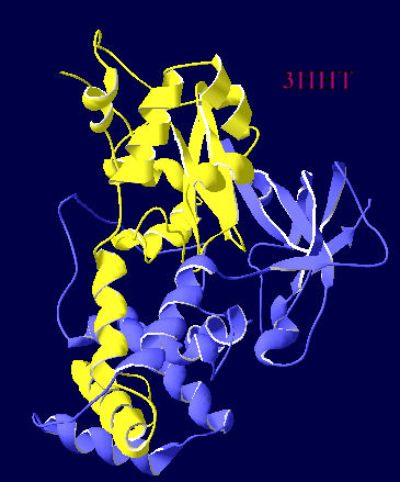
The extra helix in a cobalt-centred NHase
Using Deepview, I thought I would produce a matched pair of views of the two different metal centred NHases in ribbon view so that it was really obvious where the extra helix was. 3HHT is the crystal structure of a cobalt centred Geobacillus and 2QDY is the crystal structure of iron centred AJ270.

Subscribe to:
Posts (Atom)