Search This Blog

Showing posts with label 2QDY. Show all posts

Tuesday, 18 June 2013

New nitrile hydratase papers


Enzyme–Substrate Binding Landscapes in the Process of Nitrile Biodegradation Mediated by Nitrile Hydratase and Amidase from Yu Zhang, Zhuotong Zeng, Guangming Zeng, Xuanming Liu, Ming Chen, Lifeng Liu, Zhifeng Liu & Gengxin Xie in Applied Biochemistry and Biotechnology describes molecular modelling experiments using the crystal structures 2QDY (AJ270 Fe based NHase) and 1IRE (Pseudonocardia thermophila Co based NHase).This is a docking study for these two enzymes and a downstream amidase. Available at DOI 10.1007/s12010-013-0276-1. Shown below is Fig1b which illustrates a binding mode between the P. thermophila Co based NHase and 3-cyanopyridine.


Strategy for successful expression of the Pseudomonas putida nitrile hydratase activator P14K in Escherichia coli by Yi Liu, Wenjing Cui , Yueqin Fang, Yuechun Yu, Youtian Cui, Yuanyuan Xia, Michihiko Kobayashi and Zhemin Zhou adds to debate around the activator which is used for the maturation of the NHase with inclusion of the metal centre  (they say an activator is always needed, but is that true?). This paper is found in BMC Biotechnology at DOI:10.1186/1472-6750-13-48 and describes a methodology to ensure that the P14K activator is expressed successfully and in a more stable form.

Monday, 3 September 2012

Overlaying the NHases in 4FM4 and 2QDY

The iron centred NHase which I have most experience of is the Rhodococcus erythropolis AJ270. I have overlayed it onto the structure for the Comomonas testosteroni Ni1, and it is very similar as you might expect both at the secondary structure level (4FM4 in red, 2QDY in blue and yellow) and as an alignment. (The original paper makes in-depth analysis with 1AHJ from Rhodococcus sp. r312.)

Monday, 4 July 2011

Looking for the iron in AJ270

 Using PyMol I have been looking to see what the tunnel which connects the iron centre to the outside world looks like. Here are some pictures. Iron is represented as a warmpink sphere, and the pdb structure used is 2QDY.


Or close up as...

Wednesday, 18 May 2011

Salt bridges and hydrogen bonding

I have been playing with PISA tool as it offered insight into size and nature of the interface between the two subunits in NHases. I stuck in the PDB data for the most distinct cobalt-centred NHases- 1UGP, 1V29 and 3HHT, and PISA suggested that these have 61, 67 and 65 hydrogen bonds (respectively) between the alpha and beta subunits, and between 26 and 29 salt bridges across this interface. Compare these numbers with that for 2QDY (the iron-centred NHase from AJ270)... 51 hydrogen bonds and 8 salt bridges. That looks quite a difference... sadly things might not be so clear-cut in grouping these enzymes by metal centre- the newest PDB for 3QXE gives an average of 53 hydrogen bonds and 15 salt bridges, and that is cobalt-centred. Furthermore I do wonder, with such a small set of structures, whether we are seeing a distinction based on the fact that the first three cobalt-centred PDBs are of enzymes specifically tagged as "thermophilic", and AJ270 and Pseudomonas putida aren't. [I also looked at 2DPP and got 62 hydrogen bonds and 27 salt bridges].

Tuesday, 8 March 2011

ClustalW analysis of pdb sequences

Following on from looking at the similarity of the pdb files, I thought I would run a ClustalW analysis of the primary sequences I used for that analysis. A lot are very similar (>98%) as you might expect, and once again it is obvious these files actually represent one iron centred and two cobalt centred templates. The iron centred template has scores between 40 and 50% for the cobalt centred pair, and the score between the two cobalt centred templates is about 60%.


Monday, 7 March 2011

How many clearly different structures of NHase are reported as pdb files?

Another helpful thing that the Iterative Magic Fit function within DeepView can do is provide a numerical value (based on a RMS calculation) for the similarity of the three dimensionality of the enzymes being overlaid. After concentrating on enzymes which are clearly NHases, discarding structures which are clearly mutants of an existing structure (which is either obvious from the title or the combination of author and date of submission), you get left with 13 distinct pdb files [1AHJ, 1IRE, 1UGP, 1V29, 2AHJ, 2CYZ, 2CZ6, 2D0Q, 2DPP, 2QDY, 2ZPB, 3A8O and 3HHT]. I then IMFed every one of these against each other to get a matrix of values for similarity. Basically if they had a value of less than 0.5, they look pretty much the same, greater than 1 than they look noticeably different.
The results of this showed that ALL the iron centred enzymes have RMS of <0.5 with each other which is not entirely surprising since they are all from Rhodococcus species. They all have RMS of greater than 1 for all the cobalt centred enzymes. There are two distinctly different cobalt centred arrangements: the Pseudonocardia thermophila pair of 1IRE and IUGP, and the trio of Bacillus structures of 1V29, 2DPP and 3HHT. Within these groupings they have RMS of less than 0.5, and between the groupings RMS of greater than 1.
Effectively this says to me that there are three basic structures for NHase known currently- one iron based and two cobalt based. Below is an excerpt from a table holding all the results from these calculations which shows this clustering.
PDB ID

1.06
1.06

1.06
0.07
1.19
1.14
0.45
1.09
0.44
1.07
0.47
1.09
0.45
1.09
1.22
1.16
0.46
1.09
0.44
1.08
3A8O
0.47
1.11
3HHT
1.22
1.15

Wednesday, 2 March 2011

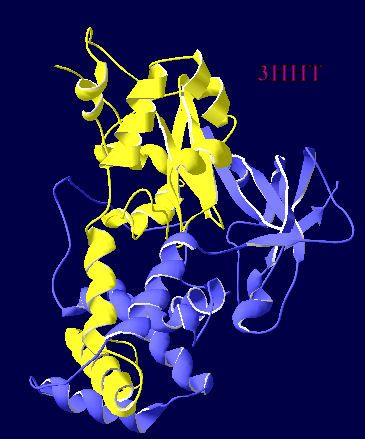
The extra helix in a cobalt-centred NHase

Using Deepview, I thought I would produce a matched pair of views of the two different metal centred NHases in ribbon view so that it was really obvious where the extra helix was. 3HHT is the crystal structure of a cobalt centred Geobacillus and 2QDY is the crystal structure of iron centred AJ270.
  

Thursday, 24 February 2011

3A8O vs 2QDY

I have been playing with Deepview which offers a handy function where you can overlay pdb files and the resulting structure is colour coded for alignment diversity. Blue indicates really not much at all. As I have mentioned before the pdb for the Rhodococcus species AJ270 is coded as 2QDY and that for Rhodococcus species N-771 is 3A8O. Overlaying these two shows almost everything is dark blue, apart from differences for the two post-translationally modified cysteines in the active site and one bit dangling off the end. So that would be very similar then. You can see the same sort of picture if you overlay these with the 2CYZ too.

Wednesday, 9 February 2011

3D structure of AJ270 NHase

We have worked with the nitrile hydratase from Rhodococcus erythropolis AJ270 which was a bacterium which was collected on the banks of the river Tyne in Newcastle. The key authors who have worked with this bacterium in synthesis over the years are Otto Meth Cohn (at Sunderland University) and Mei-Xiang Wang  (originally working with OMC but now at the Chinese Academy of Sciences, Beijing). M-X W has published a crystal structure of the nitrile hydratase from AJ270, and it can be found on PDB as 2QDY here. A little bit of playing with the data using PyMol and you can see how the two subunits fit together, and how there is a small channel from the exterior of the enzyme down into the active site.