Search This Blog

Showing posts with label AJ270. Show all posts

Tuesday, 18 June 2013

New nitrile hydratase papers


Enzyme–Substrate Binding Landscapes in the Process of Nitrile Biodegradation Mediated by Nitrile Hydratase and Amidase from Yu Zhang, Zhuotong Zeng, Guangming Zeng, Xuanming Liu, Ming Chen, Lifeng Liu, Zhifeng Liu & Gengxin Xie in Applied Biochemistry and Biotechnology describes molecular modelling experiments using the crystal structures 2QDY (AJ270 Fe based NHase) and 1IRE (Pseudonocardia thermophila Co based NHase).This is a docking study for these two enzymes and a downstream amidase. Available at DOI 10.1007/s12010-013-0276-1. Shown below is Fig1b which illustrates a binding mode between the P. thermophila Co based NHase and 3-cyanopyridine.


Strategy for successful expression of the Pseudomonas putida nitrile hydratase activator P14K in Escherichia coli by Yi Liu, Wenjing Cui , Yueqin Fang, Yuechun Yu, Youtian Cui, Yuanyuan Xia, Michihiko Kobayashi and Zhemin Zhou adds to debate around the activator which is used for the maturation of the NHase with inclusion of the metal centre  (they say an activator is always needed, but is that true?). This paper is found in BMC Biotechnology at DOI:10.1186/1472-6750-13-48 and describes a methodology to ensure that the P14K activator is expressed successfully and in a more stable form.

Wednesday, 6 July 2011

Looking inside for the active site of 3QXE

This is just a couple of rough pictures of 3QXE (the nitrile hydratase from Pseudomonas putida), once again from PyMol, showing a ribbon view of it with the active site cobalt-binding residues shown as CPK spheres, followed by a surface model with some of the active site just visible inside. I have been careful to keep it oriented similarly to the AJ270 nitrile hydratase in the previous post.

Friday, 10 June 2011

Double length equals a single subunit?

Following on from the last post... it seems reasonable to check that if you get sequences thrown up by a search which are about the double the length of one subunit in a two subunit enzyme that you don't have a situation where the two subunits are actually linked. This is the situation for the nitrile hydratase from Monosiga brevicollis which can be seen to the right of the histogram in the last post. For this reason, it seems like a good idea to check out the two sequences in the n=360 and n=380 boxes. Well the one in the n=380 box is titled "nitrile hydratase regulator [Agrobacterium radiobacter K84]" and doesn't look right at all. Scratch that one. The other one is simply called GK12401 and is from Drosophila willistoni... a eukaryote like Monosiga. Looking at the GenPept file it is easy to see how the thing was given the NHase alpha coding (I have put a green line around the bit that looks like the metal binding bit of a NHase) though I am not convinced it is what it actually is or does (based on trying to align it to Monosiga NHase or an example alpha subunit from AJ270).

Wednesday, 2 March 2011

The extra helix in a cobalt-centred NHase

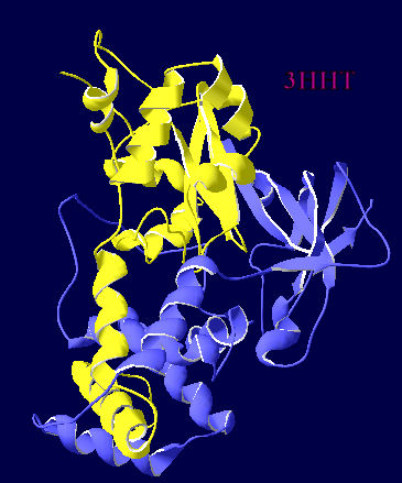
Using Deepview, I thought I would produce a matched pair of views of the two different metal centred NHases in ribbon view so that it was really obvious where the extra helix was. 3HHT is the crystal structure of a cobalt centred Geobacillus and 2QDY is the crystal structure of iron centred AJ270.
  

Saturday, 26 February 2011

Comparing 3D structures of a Fe-centred NHase and a Co-centred NHase

Using DeepView, I have used their "Iterative Magic Fit" to compare the AJ270 structure (Fe-centred) with the cobalt-centred structure reported as 3HHT by Van Wyk, Sewell, Cowan, Sayed, Tsekoa and Tastan Bishop.
The top picture shows how much homology there is between a cobalt centred NHase and an iron-centred NHase which I essentially chose at random [blue indicates similar, grading to yellow for dissimilar]. I have labelled the position of the metals in the centre of the overlayed proteins, and the text is unreadable because they fall on top of each other with this fit (which is based on aligning the carbon backbone)
The second picture is an attempt to show this metal-containing middle portion in greater detail.


This final picture above shows the 3D structure window and the primary sequence alignment window together. The lower sequence (Co-centred 3HHT) shows an extra section which isnt in the upper sequence (Fe-centred  AJ270), and corresponds to the alpha helix which is coloured yellow and lies roughly horizontally across the middle of the enzyme structure. This is a bit of structure which has been suggested as making a crucial difference in terms of extra stability.

Wednesday, 9 February 2011

3D structure of AJ270 NHase

We have worked with the nitrile hydratase from Rhodococcus erythropolis AJ270 which was a bacterium which was collected on the banks of the river Tyne in Newcastle. The key authors who have worked with this bacterium in synthesis over the years are Otto Meth Cohn (at Sunderland University) and Mei-Xiang Wang  (originally working with OMC but now at the Chinese Academy of Sciences, Beijing). M-X W has published a crystal structure of the nitrile hydratase from AJ270, and it can be found on PDB as 2QDY here. A little bit of playing with the data using PyMol and you can see how the two subunits fit together, and how there is a small channel from the exterior of the enzyme down into the active site.