Search This Blog

Showing posts with label 3HHT. Show all posts

Friday, 3 August 2012

NHase in halophilic archaeon DL31

I notice that both an alpha chain and a beta chain of a cobalt centred NHase have been recently annotated (14 June 2012) in the genome of the rather unprepossessingly named "halophilic archaeon DL31". It must be quite an elusive beast because Google Images doesn't deliver a picture of anything suitable.
Halophilicity quite often comes with tolerance to alkaline conditions, which can mean all sorts of utility in the world of synthetic biocatalysis. As I mentioned in the "No extremes" post we published with Roger Sheldon's group on the synthetic potential of one of these halophilic NHases, but this is a good example of a genome-sourced potential halophilic NHase. BLASTing both the alpha and beta chains against prokaryotic genomes shows that the beta chain is most similar to beta chain of Caldalkalibacillus thermarum TA2.A1 (I think we can guess the proclivities of this microbe) with 3 of the next 4 best hits being the sequences from the crystal structures 3HHT, 2DPP and 1V29 all of which are moderately thermophilic.


Interestingly there is a slightly different aspect to the alpha chain BLAST: Pseudonocardia dioxanivorans and Rhodopseudomonas palustris make up the top hits before Caldalkalibacillus thermarum TA2.A1 turns up.

Wednesday, 6 July 2011

Comparing the access of the active site of 3QXE and 3HHT

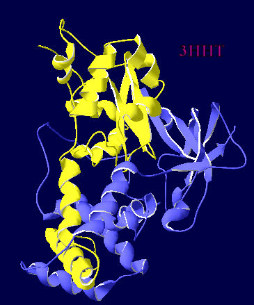
If you orient the pdb file of the Geobacillus which is coded as 3HHT as I have done for 3QXE and 2QDY in the previous posts, you get the following two images.


The cobalt is no longer visible. However if you twirl the structure around you can find a view of it through a different aperture. I wonder how this impacts on the selectivity of this enzyme.


Wednesday, 18 May 2011

Salt bridges and hydrogen bonding

I have been playing with PISA tool as it offered insight into size and nature of the interface between the two subunits in NHases. I stuck in the PDB data for the most distinct cobalt-centred NHases- 1UGP, 1V29 and 3HHT, and PISA suggested that these have 61, 67 and 65 hydrogen bonds (respectively) between the alpha and beta subunits, and between 26 and 29 salt bridges across this interface. Compare these numbers with that for 2QDY (the iron-centred NHase from AJ270)... 51 hydrogen bonds and 8 salt bridges. That looks quite a difference... sadly things might not be so clear-cut in grouping these enzymes by metal centre- the newest PDB for 3QXE gives an average of 53 hydrogen bonds and 15 salt bridges, and that is cobalt-centred. Furthermore I do wonder, with such a small set of structures, whether we are seeing a distinction based on the fact that the first three cobalt-centred PDBs are of enzymes specifically tagged as "thermophilic", and AJ270 and Pseudomonas putida aren't. [I also looked at 2DPP and got 62 hydrogen bonds and 27 salt bridges].

Tuesday, 8 March 2011

ClustalW analysis of pdb sequences

Following on from looking at the similarity of the pdb files, I thought I would run a ClustalW analysis of the primary sequences I used for that analysis. A lot are very similar (>98%) as you might expect, and once again it is obvious these files actually represent one iron centred and two cobalt centred templates. The iron centred template has scores between 40 and 50% for the cobalt centred pair, and the score between the two cobalt centred templates is about 60%.


Monday, 7 March 2011

How many clearly different structures of NHase are reported as pdb files?

Another helpful thing that the Iterative Magic Fit function within DeepView can do is provide a numerical value (based on a RMS calculation) for the similarity of the three dimensionality of the enzymes being overlaid. After concentrating on enzymes which are clearly NHases, discarding structures which are clearly mutants of an existing structure (which is either obvious from the title or the combination of author and date of submission), you get left with 13 distinct pdb files [1AHJ, 1IRE, 1UGP, 1V29, 2AHJ, 2CYZ, 2CZ6, 2D0Q, 2DPP, 2QDY, 2ZPB, 3A8O and 3HHT]. I then IMFed every one of these against each other to get a matrix of values for similarity. Basically if they had a value of less than 0.5, they look pretty much the same, greater than 1 than they look noticeably different.
The results of this showed that ALL the iron centred enzymes have RMS of <0.5 with each other which is not entirely surprising since they are all from Rhodococcus species. They all have RMS of greater than 1 for all the cobalt centred enzymes. There are two distinctly different cobalt centred arrangements: the Pseudonocardia thermophila pair of 1IRE and IUGP, and the trio of Bacillus structures of 1V29, 2DPP and 3HHT. Within these groupings they have RMS of less than 0.5, and between the groupings RMS of greater than 1.
Effectively this says to me that there are three basic structures for NHase known currently- one iron based and two cobalt based. Below is an excerpt from a table holding all the results from these calculations which shows this clustering.
PDB ID

1.06
1.06

1.06
0.07
1.19
1.14
0.45
1.09
0.44
1.07
0.47
1.09
0.45
1.09
1.22
1.16
0.46
1.09
0.44
1.08
3A8O
0.47
1.11
3HHT
1.22
1.15

Wednesday, 2 March 2011

The extra helix in a cobalt-centred NHase

Using Deepview, I thought I would produce a matched pair of views of the two different metal centred NHases in ribbon view so that it was really obvious where the extra helix was. 3HHT is the crystal structure of a cobalt centred Geobacillus and 2QDY is the crystal structure of iron centred AJ270.
  

Saturday, 26 February 2011

Comparing 3D structures of a Fe-centred NHase and a Co-centred NHase

Using DeepView, I have used their "Iterative Magic Fit" to compare the AJ270 structure (Fe-centred) with the cobalt-centred structure reported as 3HHT by Van Wyk, Sewell, Cowan, Sayed, Tsekoa and Tastan Bishop.
The top picture shows how much homology there is between a cobalt centred NHase and an iron-centred NHase which I essentially chose at random [blue indicates similar, grading to yellow for dissimilar]. I have labelled the position of the metals in the centre of the overlayed proteins, and the text is unreadable because they fall on top of each other with this fit (which is based on aligning the carbon backbone)
The second picture is an attempt to show this metal-containing middle portion in greater detail.


This final picture above shows the 3D structure window and the primary sequence alignment window together. The lower sequence (Co-centred 3HHT) shows an extra section which isnt in the upper sequence (Fe-centred  AJ270), and corresponds to the alpha helix which is coloured yellow and lies roughly horizontally across the middle of the enzyme structure. This is a bit of structure which has been suggested as making a crucial difference in terms of extra stability.